Co-reporter:Masafumi Tanaka, Akira Hosotani, Yuka Tachibana, Minoru Nakano, Kenji Iwasaki, Toru Kawakami, and Takahiro Mukai
Langmuir 2015 Volume 31(Issue 46) pp:12719-12726
Publication Date(Web):November 4, 2015
DOI:10.1021/acs.langmuir.5b03438
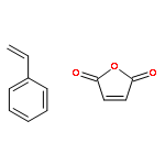
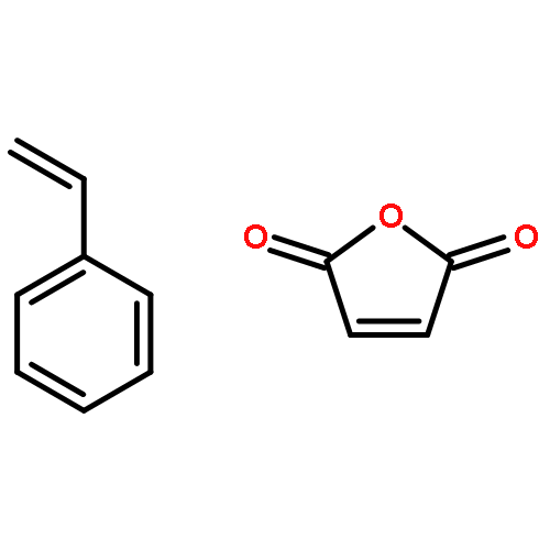
Discoidal high-density lipoproteins generated by the apolipoprotein-mediated solubilization of membrane lipids in vivo can be reconstituted with phospholipids and apolipoproteins in vitro. Recently, it has been reported that such particles can be prepared using the hydrolyzed acid form of styrene–maleic anhydride copolymer (SMAaf) instead of apolipoproteins, but characterization of its physicochemical properties has remained less elucidated. In the present study, with the aim of applying SMAaf-based lipid nanoparticles as novel delivery vehicles of drugs and/or imaging agents, we investigated the preparation conditions and evaluated the physicochemical properties of lipid–SMAaf complexes. SMAaf induced spontaneous turbidity clearance of dimyristoylphosphatidylcholine (DMPC) vesicles accompanied by the formation of smaller particles not only at the phase transition temperature of DMPC but also above it. Such reductions in the turbidity were not observed with some other amphiphilic synthetic polymers tested under the same experimental conditions. Size exclusion chromatography analyses showed that homogeneously sized particles were prepared at lipid to SMAaf weight ratios of less than 1/1.5. Dynamic light scattering and transmission electron microscopy revealed that gel-filtered DMPC–SMAaf complexes were approximately 8–10 nm in diameter and discoidal in shape. The DMPC–SMAaf complexes were relatively stable even after lyophilization but were sensitive to pH changes. Fluorescence techniques demonstrated that the gel to liquid-crystalline phase transition temperature of DMPC in the discoidal complexes broadened significantly relative to that of liposomes, despite their common bilayer structure, which is a typical feature of discoidal lipid nanoparticles. These results provide fundamental insights into discoidal SMAaf-based lipid nanoparticles for the development of novel delivery vehicles.
Co-reporter:Shinya Ohta, Masafumi Tanaka, Kota Sakakura, Toru Kawakami, Saburo Aimoto, Hiroyuki Saito
Chemistry and Physics of Lipids 2009 Volume 162(1–2) pp:62-68
Publication Date(Web):November 2009
DOI:10.1016/j.chemphyslip.2009.07.008
Human serum amyloid A (SAA) protein is an apolipoprotein predominantly present in the high-density lipoprotein fraction of plasma. Despite its critical roles in lipid metabolism, especially in acute phases, systematic understanding of the lipid interaction of this protein is limited. Lipid-binding properties of synthetic fragment peptides corresponding to the N-terminal (residues 1–27), central (residues 43–63), and C-terminal (residues 77–104) parts of SAA molecule were examined. SAA (1–27) peptide binds to lipid forming an α-helical structure, whereas SAA (43–63) and (77–104) peptides do not display binding to lipid with any conformational changes. These results indicate that the N-terminal region of SAA is important for lipid interaction. In addition, the finding that deletion of or proline substitution in the most N-terminal region (residues 1–11) markedly decreased the binding to lipid further suggests that the α-helical structure in residues 1–11 is essential for lipid binding of SAA.
Co-reporter:Hiroka Takase, Hiroki Furuchi, Masafumi Tanaka, Toshiyuki Yamada, Kyoko Matoba, Kenji Iwasaki, Toru Kawakami, Takahiro Mukai
Biochimica et Biophysica Acta (BBA) - Molecular and Cell Biology of Lipids (October 2014) Volume 1841(Issue 10) pp:
Publication Date(Web):October 2014
DOI:10.1016/j.bbalip.2014.07.012
•α-Helix formation was induced upon binding of SAA to phospholipid vesicles at 37 °C.•SAA formed rHDL particles with the size of native HDL by phospholipid solubilization.•Lipid binding rendered SAA molecules resistant to protein degradation.•Physicochemical properties of rHDL reconstituted from three SAA isoforms were similar.•The present study provides fundamental HDL models under inflammatory conditions.The acute-phase human protein serum amyloid A (SAA) is enriched in high-density lipoprotein (HDL) in patients with inflammatory diseases. Compared with normal HDL containing apolipoprotein A-I, which is the principal protein component, characteristics of acute-phase HDL containing SAA remain largely undefined. In the present study, we examined the physicochemical properties of reconstituted HDL (rHDL) particles formed by lipid interactions with SAA. Fluorescence and circular dichroism measurements revealed that although SAA was unstructured at physiological temperature, α-helix formation was induced upon binding to phospholipid vesicles. SAA also formed rHDL particles by solubilizing phospholipid vesicles through mechanisms that are common to other exchangeable apolipoproteins. Dynamic light scattering and nondenaturing gradient gel electrophoresis analyses of rHDL after gel filtration revealed particle sizes of approximately 10 nm, and a discoidal shape was verified by transmission electron microscopy. Thermal denaturation experiments indicated that SAA molecules in rHDL retained α-helical conformations at 37 °C, but were almost completely denatured around 60 °C. Furthermore, trypsin digestion experiments showed that lipid binding rendered SAA molecules resistant to protein degradation. In humans, three major SAA1 isoforms (SAA1.1, 1.3, and 1.5) are known. Although these isoforms have different amino acids at residues 52 and 57, no major differences in physicochemical properties between rHDL particles resulting from lipid interactions with SAA isoforms have been found. The present data provide useful insights into the effects of SAA enrichment on the physicochemical properties of HDL.
Co-reporter:Masashi Egashira, Hiroka Takase, Izumi Yamamoto, Masafumi Tanaka, Hiroyuki Saito
Archives of Biochemistry and Biophysics (July 2011) Volume 511(Issues 1–2) pp:101-106
Publication Date(Web):July 2011
DOI:10.1016/j.abb.2011.04.019
Co-reporter:Masafumi Tanaka, Ayaka Nishimura, Haruka Takeshita, Hiroka Takase, Toshiyuki Yamada, Takahiro Mukai
Chemistry and Physics of Lipids (January 2017) Volume 202() pp:
Publication Date(Web):January 2017
DOI:10.1016/j.chemphyslip.2016.11.004
•Amyloid fibril formation of human serum amyloid A (SAA) under a lipid environment was investigated.•SAA (1–27) peptide bound to neutral and acidic, whereas SAA (43–63) peptide bound only to acidic lysophospholipids.•For both these SAA peptides, binding to lysophospholipids inhibited heparin-induced amyloid-like fibril formation.•Acidic lysophospholipids implied a possibility to promote fibril formation of SAA (1–27) peptide by themselves.•Amyloid fibril formation of SAA may be modulated by altering the lipid head group composition of HDLs during metabolism.Human serum amyloid A (SAA) is a precursor protein of AA amyloidosis and a component of high-density lipoproteins (HDLs), thus it is essential to investigate the amyloid fibril formation of SAA under a lipid environment. We used synthetic fragment peptides corresponding to the N-terminal (residues 1–27) and central (residues 43–63) regions of the SAA molecule, which are known to have amyloidogenic properties. Measurements of tryptophan fluorescence in conjunction with circular dichroism showed that SAA (1–27) peptide binds to neutral and acidic lysophospholipids, whereas SAA (43–63) peptide binds only to acidic lysophospholipids. For both these SAA peptides, binding to lysophospholipids inhibited heparin-induced amyloid-like fibril formation by stabilizing the α-helical structure. However, acidic lysophospholipids implied a possibility to promote fibril formation of SAA (1–27) peptide by themselves. These results suggest that the amyloid fibril formation of SAA may be modulated by altering the lipid head group composition of HDLs during metabolism.
Co-reporter:Masafumi Tanaka, Padmaja Dhanasekaran, David Nguyen, Margaret Nickel, Yuki Takechi, Sissel Lund-Katz, Michael C. Phillips, Hiroyuki Saito
Biochimica et Biophysica Acta (BBA) - Molecular and Cell Biology of Lipids (January 2011) Volume 1811(Issue 1) pp:
Publication Date(Web):January 2011
DOI:10.1016/j.bbalip.2010.10.003
As the principal component of high-density lipoprotein (HDL), apolipoprotein (apo) A-I plays essential roles in lipid transport and metabolism. Because of its intrinsic conformational plasticity and flexibility, the molecular details of the tertiary structure of lipid-free apoA-I have not been fully elucidated. Previously, we demonstrated that the stability of the N-terminal helix bundle structure is modulated by proline substitution at the most hydrophobic region (residues around Y18) in the N-terminal domain. Here we examine the effect of proline substitution at S55 located in another relatively hydrophobic region compared to most of the helix bundle domain to elucidate the influences on the helix bundle structure and lipid interaction. Fluorescence measurements revealed that the S55P mutation had a modest effect on the stability of the bundle structure, indicating that residues around S55 are not pivotally involved in the helix bundle formation, in contrast to the insertion of proline at position 18. Although truncation of the C-terminal domain (Δ190–243) diminishes the lipid binding of apoA-I molecule, the mutation S55P in addition to the C-terminal truncation (S55P/Δ190–243) restored the lipid binding, suggesting that the S55P mutation causes a partial unfolding of the helix bundle to facilitate lipid binding. Furthermore, additional proline substitution at Y18 (Y18P/S55P/Δ190–243), which leads to a drastic unfolding of the helix bundle structure, yielded a greater lipid binding ability. Thus, proline substitutions in the N-terminal domain of apoA-I that destabilized the helix bundle promoted lipid solubilization. These results suggest that not only the hydrophobic C-terminal helical domain but also the stability of the N-terminal helix bundle in apoA-I are important modulators of the spontaneous solubilization of membrane lipids by apoA-I, a process that leads to the generation of nascent HDL particles.Research Highlights►S55P mutation had a modest effect on the stability of the bundle structure of apoA-I. ►S55P/Δ190–243 apoA-I restored the lipid binding despite the C-terminal truncation. ►Destabilized N-terminal helix bundle promoted lipid solubilization. ►Alterations in the stability of apoA-I appear to influence the production of HDL.
Co-reporter:Masafumi Tanaka, Yuki Takamura, Toru Kawakami, Saburo Aimoto, ... Takahiro Mukai
FEBS Letters (1 March 2013) Volume 587(Issue 5) pp:510-515
Publication Date(Web):1 March 2013
DOI:10.1016/j.febslet.2013.01.026
Amphipathic helix, which senses membrane curvature, is of growing interest. Here we explore the effect of amino acid distribution of amphipathic helical peptide derived from the C-terminal region (residues 220–241) of human apolipoprotein (apo) A-I on membrane curvature sensing. This peptide preferred a curved membrane in a manner similar to full-length apoA-I, although its model peptide did not sense membrane curvature. Substitution of several residues both on the polar and non-polar faces of the amphipathic helix had no significant effect on sensing, suggestive of the elaborate molecular architecture in the C-terminal helical region of apoA-I to exert lipid efflux function.Highlights► The C-terminal peptide (residues 220–241) of human apoA-I preferred curved membrane. ► In contrast, its model peptide did not sense membrane curvature. ► Substitution of several amino acid residues had no significant effect on the sensing. ► The C-terminal region of apoA-I is finely designed to recognize membrane curvature.

![3,5,8-Trioxa-4-phosphahexacos-17-en-1-aminium,4-hydroxy-N,N,N-trimethyl-9-oxo-7-[[(1-oxohexadecyl)oxy]methyl]-, inner salt,4-oxide, (7R,17Z)-](http://img.cochemist.com/ccimg/26900/26853-31-6.png)
![3,5,8-Trioxa-4-phosphahexacos-17-en-1-aminium,4-hydroxy-N,N,N-trimethyl-9-oxo-7-[[(1-oxohexadecyl)oxy]methyl]-, inner salt,4-oxide, (7R,17Z)-](http://img.cochemist.com/ccimg/26900/26853-31-6_b.png)


![(6R,9AR)-OCTAHYDRO-2H-PYRIDO[1,2-A]PYRAZIN-6-YLMETHANOL](http://img.cochemist.com/ccimg/13700/13699-48-4.png)
![(6R,9AR)-OCTAHYDRO-2H-PYRIDO[1,2-A]PYRAZIN-6-YLMETHANOL](http://img.cochemist.com/ccimg/13700/13699-48-4_b.png)